
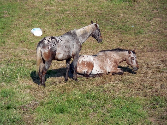
Well I am sure you have heard the phrase, “you look like you’re asleep on your feet,” it has been said to me a few times, but unfortunately it’s not possible for us humans without falling over. The cool thing and the Fun Fact about our 4 legged Equine Buddies is that they can sleep on their feet, quite literally!
Being a plains grazer the horse’s evolution has allowed them to develop a special mechanism, called The Stay Apparatus of the Horse, whereby a series of muscles, tendons and ligaments of the both the fore and hind limbs, will lock the animal’s limbs in place will minimal expenditure of energy, allowing the horse to sleep while standing. How Amazing is that?? This is useful trick if there are any predators around, because it affords the horse a speedy get away when and if he is attacked.
That being said, let’s explore what muscles, tendons and ligaments that are involved starting with the Stay Apparatus of the Forelimb.
The Stay Apparatus of the Forelimb
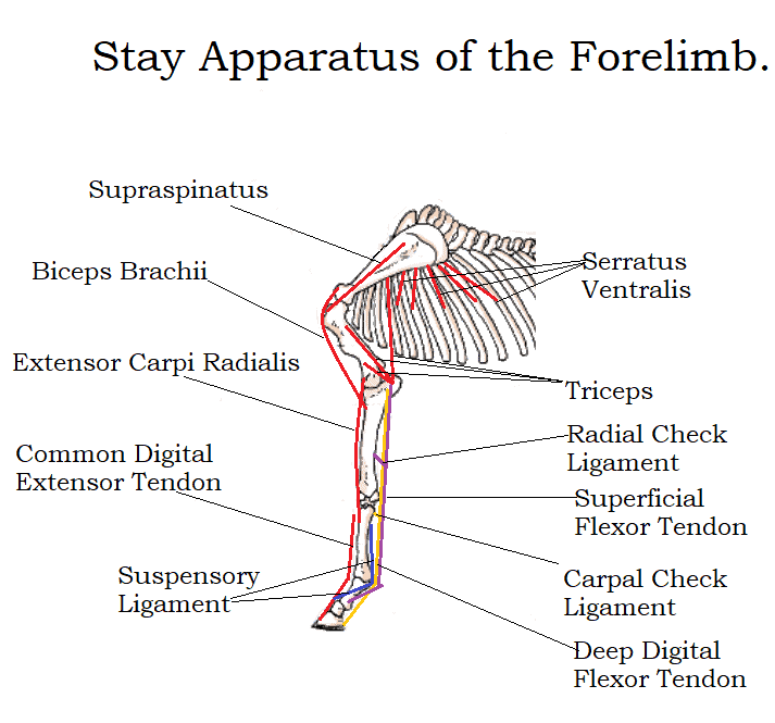
The Upper part of the forelimb, both the Shoulder and Elbow Joint are locked in place by a collaboration of the muscles. Toward the front of the limb The Supraspinatus isometrically contracts to prevent flexion at the shoulder. This Job is aided by the Biceps Brachii Muscle and the Extensor Carpi Radialis to the front of the limb. Toward the back of the Limb, the Serratus Ventralis helps to prevent extension of the Shoulder Joint, as does the Long Head of the Triceps which as it inserts into to the elbow and prevents Flexion at the Elbow Joint. The combined efforts of these muscles work in harmony to lock both the shoulder and Elbow Joints in place while the horse is sleeping while standing.
The Lower part of the forelimb and its fixation is aided by the shape of the of limb and the collaboration of the Extensor Tendons running down the front of the limb, along with the tendons of the Superficial and Deep Flexor Tendons running down the back of the limb. The Radial Check Ligament above the Knee that inserts into the Superficial Flexor tendon and the Carpal Check ligament below the knee which inserts in to the Deep Digital Flexor tendon, help to lock the knee joint in place. The normal standing position causes tension in the suspensory ligament, which is engaged to prevent over extension of the fetlock joint, this ligament splits in two at distal aspect of the fetlock joint inserting into 2 sesamoid bones, before the ligaments continues down and forward to insert into the extensor tendons on the front of the limb, aiding in keeping the extensor tendons held in position.
Now Lets Take a Look at the Stay Apparatus of the Hind Limb and Reciprocal System.
The Upper Part of the Hind Limb
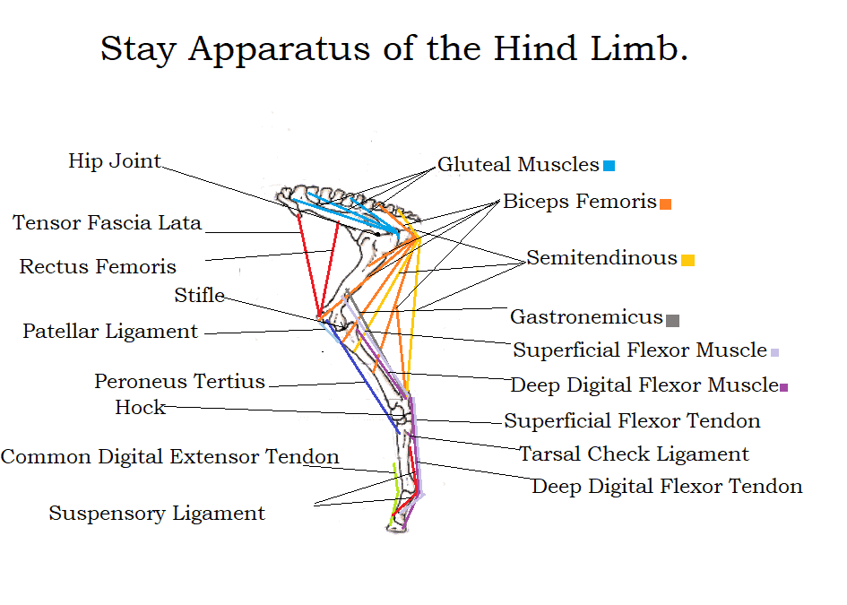
As the horse falls asleep while standing he will normally rest one hind limb with the toe on the ground, this initiates the stay apparatus of the opposite hind limb. The Reciprocal System of the hind limb of the horse, refers to that of the hock and stifle joints, which are designed to work in unison, i.e. when flexion occurs at the stifle, flexion also occurs at the hock and the same goes for extension of either. This arrangement is brought about by the peroneus tertius tendon on the front of the hind limb, which runs from the lower femur and inserts into the front of the hind cannon bone (PT normally a hock flexor) and the Gastronemicus (G normally a hock extensor) and the superficial flexor tendon on the back of the limb, which run from the lower femur and insert into the tuber calcis or point of hock. The opposing action of this reciprocal system, allows for flexion and extension of the stifle and hock to be in unison while aiding in the stabilization of the stifle and hock joints when the limb is firmly planted on the ground.
The patella is locked into place by the medial patellar ligament when the limb is weight bearing, this is helped by the action of the biceps fermoris which helps to pull the patella up the trochlear groove of the femur where it is locked by the medial patellar ligament, the rectus femoris muscle, and tensor fascia lata play a role in the stabilization of the stifle joint, while the semitendinous and the Gluteal Muscles also play a role in stabilizing the back of the limb.
The Lower Part of the Hind Limb
The stay apparatus of the lower hind limb from the hock down, works almost identically to that of the forelimb. The only difference is that the deep digital flexor tendon which receives a check ligament below the hock joint. As in the forelimb the arrangement of the extensor tendons to the front of the limb and the superficial and deep digital flexor tendons to the back of the limb, along with the suspensory ligament, help to fix and support the joints of the lower hind limb.
Quite a complicated set up by all accounts, that happens in the blink of an eye as your favourite equine grabs forty winks on a warm sunny day.
This Fun Horse Fact was originally published in the February 2021 Issue of Irish Sport Horse magazine – Anatomy Pictures are the Property of Irish Sport Horse Magazine.